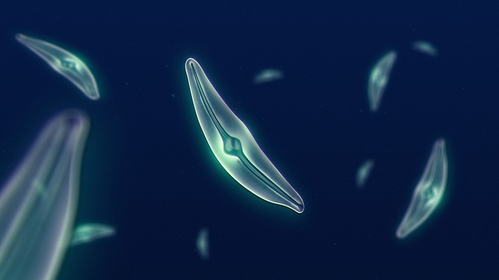
In this article we will discuss about:- 1. Description of Bacillariophyceae 2. Characteristics of Bacillariophyceae 3. Occurrence 4. Cell Structure 5. Reproduction 6. Economic Importance.
1. Description of Bacillariophyceae
It is a large group of algae consisting of 200 genera and over 10,000 species, out of which 92 genera and about 569 species are reported from India. They are commonly known as Diatoms. The diatoms are the most beautiful microscopic algae due to their structure and sculpturing of their walls.
2. Characteristics of Bacillariophyceae
2. Microscopic cells are of different shapes. They may be oval, spherical, triangular, boat- shaped etc.
3. Plant bodies are either bilateral or radial in symmetry.
4. The cells are surrounded by a rigid cell wall, called frustule, consisting of upper epitheca and lower hypotheca; arranged in the form of a box with its lid.
5. The cell wall is composed of pectic substances impregnated with high amount of siliceous substance.
6. The wall may have secondary structures like spines, bristles etc.
7. Vegetative cells are diploid (2n).
8. The cells generally have many discoid or two large plate-like chromatophores. Some cells possess stellate chromatophore.
9. The photosynthetic pigments are chlorophyll a, chlorophyll c along with xanthophylls like fucoxanthin, diatoxanthin and diadinoxanthin.
10. Reserve food is oil, volutin and crysolaminarin.
11. Some vegetative cells show gliding movement.
12. Motile structure (antherozoid) has single pantonematic flagellum.
13. Vegetative multiplication takes place by cell division, which is very common. Some of the cells become very much reduced in size.
14. They produce characteristic spore, the auxospore which develops to regain the normal size.
15. Sexual reproduction takes place by isogamy and oogamy.
3. Occurrence
The terrestrial species (Amorpha, Navicula, Pinnularia etc.) are able to withstand desiccation for a long period. Some diatoms (Gomphonima, Cymbella etc.) can grow as epiphyte on other algae (Enteromorpha, Cladophora etc.) and higher plant. Licmophora, a member of diatom, grows endozoically.
4. Cell Structure
Both the theca consist of two portions:
(a) Valve — the upper flattened top and
(b) Connecting band or cingulum (pl. cingula) — the incurved region.
The common region of the connecting bands, where both the theca remain fitted together, is the girdle. [When the diatoms are observed from the valve side i.e., valve side is uppermost, called the valve view, but when viewed from the connecting band, it is the girdle view]. Depending on symmetry, the cells are divided into two orders: Pennales (bilaterally symmetry) and Centrales (radially symmetry).
In some pinnate diatoms (Cybella cistula, Pinnularia viridis etc.) an elongated slit is present on their valves, called raphe. The raphe is interrupted at its midpoint by thickening of the wall called central nodule. Similar thickening is also present at the ends called polar nodules. Some members like Tabellaria fenestrate etc. of the order Pennales, do not have raphe, called pseudoraphe.
Besides raphe or pseudoraphe, the cell walls have other types of openings, called pores and locules.
Based on electron microscopic studies, Hendey (1971) observed four basic types of secondary structures. These are: Punctae (small perforations on valve surface), Canaliculi (tubelike narrow channels which run through the valve surface), Areolae (large boxlike depressions) and Costae (riblike structures on the valve surface).
The cell wall is mainly made up of pectic substances, impregnated with silica. The content of silica varies from 1% (Phaeodactylum tricornutum) to about 50% on the basis of dry weight of the cell.
Protoplast:
The entire content present inside the cell wall is the protoplast. The cell membrane encloses a large central vacuole surrounded by cytoplasm. The cytoplasm contains single nucleus, mitochondria, golgi bodies and chloroplasts. The chloroplasts may be of different shapes like stellate, H-shaped, discoid etc. In some species the chloroplasts contain pyrenoids.
The photosynthetic pigments are chlorophyll a, c1 and c2, β-carotene, fucoxanthin, diatoxanthin and diadinoxanthin. The latter two are present in small quantity. (The golden-brown colour of diatom cells is due to the presence of xanthophylls like fucoxanthin, diatoxanthin and diadinoxanthin.
The term diatomin is used for the mixture of chlorophyll and carotenoids, particularly carotene and several brown xanthophylls pigments.) The reserve food of diatoms is chrysolami- narin and oil droplets (they do not store in the form of starch).
Locomotion:
All diatoms with raphe are motile. Most of the members of the order Pennales contain raphe and perform gliding movement. The gliding movement is caused by the circulation of cytoplasm within the raphe by the release of mucilage. The rate of movement varies from 02-25 µm/sec. The locomotion is affected by temperature, light etc
ggggggggggggggg
5. Reproduction
1. Vegetative Reproduction:
Vegetative reproduction performs with the help of cell division (Fig. 3.102). It takes place usually at midnight or in the early morning.
During cell division the protoplast of the cell enlarges slightly, thus the cell increases in volume and slightly separates both the theca (epitheca and hypotheca). Then the protoplast undergoes mitotic division and gets separated along the longitudinal axis through the median line.
Thus one half of protoplast remains in epitheca and the other one in hypotheca. One side of the protoplast thus remains naked. Now both the theca i.e., epitheca and hypotheca of mother cell behave as epitheca of the daughter cells.
Thus new silicious valves are deposited towards the naked sides of the protoplast and always behave as hypotheca of the daughter cells. Connecting bands are developed between the theca. Later on, the daughter cells get separated.
During cell division, both the theca i.e., epitheca and hypotheca of the mother cell behave as epitheca of the daughter cells. So at the side where the hypotheca behaves as epitheca, the cell becomes reduced in size. Thus with continuous cell division some cells gradually become reduced in size.
2. Sexual Reproduction:
The pattern of sexual reproduction differs in both orders — Pennales and Centrales. During this process, auxospore is formed in both the groups. During cell division, those cells become reduced in size, are able to regain their normal size through the formation of auxospore, so it is a “restorative process” rather than multiplication.
Auxospore Formation in Pennales:
It takes place through gametic union, autogamy and parthenogenesis.
These are of the following types:
1. Production of one auxospores by two conjugating cells. In this process two uniting cells come very close to each other (Fig. 3.103) and become covered by a mucilaginous sheath. The diploid nucleus of each cell undergoes meiosis.
Out of four nuclei, three degenerate and only one survives. The surviving nucleus behaves as gamete (n). The gametes come out from the parent frustules and unite together, to form a zygote (2n).
After a short period of rest the zygote elongates considerably and functions as an auxospore. The auxospore projects out from the parent frustules along with mucilage and elongates in a plane parallel to the long axis of the parent diatom.
The auxospore is enclosed in a pectic membrane, the perizonium. The auxospore then develops new frustule inside the perizonium. Thus new diatom cell is formed which regains the normal size. It is found in Cocconis placentula, Surirella saxonica etc.
2. Production of Two Auxospores by Two Conjugating Cells:
This is a very common process of auxospore formation. In this process the conjugating cells come very close to each other and get enclosed by mucilage (Fig. 3.104). The nucleus (2n) of each cell undergoes meiotic division and forms four nuclei.
Out of four nuclei, two degenerate, the rest two survive. The cytoplasm then divides either equally or unequally and along with one nucleus they behave as gametes. Thus two gametes are formed in each cell.
The pattern of union between the gametes varies from species to species. Both the gametes of a cell may be active and fuse with the gametes of other cell, thus two zygotes are produced in a single cell or out of two, one becomes active and fertilises with the opposite one and thus one zygote is produced in each cell.
The zygotes elongate and function as auxo- spores. The auxospores develop the perizonium around themselves and both of them develop new frustules on their outer sides i.e., inside the perizonium. Thus two diatom cells of normal size are formed. It is found in Cymbella lanceolata, Gomphomema parvulum etc.
3. Production of One Auxospore by One Cell:
This process of auxospore formation is called Paedogamy (Pedogamy). In this process the diploid nuclei of a vegetative cell undergo meiosis and form four haploid nuclei. Out of the four nuclei two partially degenerate. Each of the rest two along with the cytoplasm and one partially degenerated nucleus, behaves as gamete. Later on, the union between the two sister gametes takes place and forms the zygote.
The zygote comes out from the parent frustule and behaves as an auxospore. The auxospore then gets covered by perizonium and develops wall inside the perizonium. Thus one diatom cell of normal size is formed.
4. Production of One Auxospore by Autogamy:
In this process the diploid nucleus undergoes first meiotic division. Thus two haploid nuclei are formed. The two nyclei in the protoplast come side by side, fuse together and form diploid (2n) nucleus. This is called autogamous pairing.
The protoplast along with diploid (2n) nucleus comes out from the parent frustule and behaves as an auxospore. The auxospores are then covered by perizonium. New wall develops on the auxospore inner to the perizonium. Thus a new individual of normal size is developed. This is found in Amphora normani.
5. Production of Auxospore by Parthenogenesis:
The diatom cells come together and are covered by a common mucilage envelop (Fig. 3.105). The diploid nucleus undergoes two sequential mitotic divisions. Meiotic division does not take place here. One nucleus in each mitotic division degenerates. Thus only one diploid (2n) nucleus along with protoplast remains, and comes out from the mother cell and behaves as an auxospore.
The auxospore is then covered by perizonium and secretes new wall around itself. Thus normal size cell is formed.
6. Production of Auxospore by Oogamy:
In this process (Fig. 3.106) the nucleus (2n) of female cell which behaves as oogonium, undergoes meiosis and forms four nuclei. The protoplast is also divided into two unequal parts, each containing two nuclei.
The lower half is larger and behaves as functional ovum and the upper smaller one as non-functional ovum. The functional ovum contains one functional nucleus and one non-functional nucleus, which gradually degenerates at maturity.
The male cell (2n) behaves as antheridium, also undergoes meiosis and forms four nuclei. The protoplast also divides into two parts. Thus two microgametes are formed. Each of which contains two nuclei, of which one is functional and other is non-functional. The microgametes are naked, globular and non-flagellate.
After coming out, the male gamete fertilizes the egg and forms the zygote (2n). Later it functions as an auxospore and forms new individual of normal size. It is found in Rhabdonema adriaticum.
Auxospore Formation in Centrales
It takes place by autogamy and oogamy:
1. Auxospore Formation by Autogamy:
The protoplast of the vegetative cell (Fig. 3.107) secretes mucilage which separates both the theca. The nucleus (2n) then undergoes meiosis and forms four nuclei. Of the four nuclei two degenerate and the other two undergo fusion to form diploid (2n) nucleus again.
This is called autogamy. The protoplast with 2n nucleus functions as an auxospore. The auxospore forms fresh frustule inside the perizonium covering and forms cell of normal size. It is found in Melosira nummuloides.
2. Auxospore Formation by Oogamy:
Oogamy takes place by the fusion of egg and sperm developed inside the oogonium and antheridium respectively (Fig. 3.108).
Oogonium:
Single vegetative cell behaves as an oogonium. The protoplast of oogonium undergoes meiotic division and forms four nuclei. Of the four nuclei three degenerate and the remaining one functions as an egg.
Antheridium:
The pattern of development of sperms varies in different species. In species like Melosira varians the protoplast undergoes meiotic division and forms four haploid nuclei. Each haploid nucleus with some protoplast metamorphoses into an uniflagellate (tinsel type) sperm. In others the number of sperms may go up to 8 or even 128.
Fertilisation:
After coming out of the antheridium only one sperm enters inside the oogonium and fertilises the egg. The resultant zygote undergoes mitotic division but one nucleus degenerates in each division. The remaining nucleus with its protoplast behaves as an auxospore. The auxospore then develops new wall inside the perizonium covering and forms new cell of normal size like the mother. It is also called firstling cell.
From the above processes of sexual reproduction in both pennales and centrales, it becomes clear that the sexual process in diatom does not lead to multiplication but is to regain the normal size.
3. Resting Spores:
These spores are formed during unfavourable conditions. Some members reproduce by the formation of thick-walled resting spores, the cysts or statospores. They are formed in Melosira.
6. Economic Importance.
The diatoms are used in various purposes either directly or indirectly.
The different uses of diatoms are:
1. Diatomite:
After the death of diatom cells the outer coverings i.e., the silicified walls become accumulated at the bottom of water. The accumulation may be thicker during favourable conditions. These deposits are called diatomaceous earth, diatomite or keiselghur.
It is very suitable for use in different industries:
a. As Filter:
It is used as filter in different industries like sugar (to filter microorganism), oil and chemical industry. Diatomite is also used as filter for battery boxes.
b. As Insulator:
It is used as insulator in boilers and blast furnaces for its heat- resistant ability.
c. As Absorbent:
It is used as absorbent of liquid nitroglycerine.
d. Other Uses:
Diatomite is used as abrasive (i.e., capable of rubbing or grinding down) substance for the manufacture of metal paints, polish, varnish, toothpaste etc. It is also used with bake-lite for electrical fuse and switch boxes.
2. Petroleum:
Much of the petroleum is considered to be of diatom origin as they are found in association with large oil deposits.
3. Food:
Due to their great abundance in the different seas and their use as food by marine animals, they are called the ‘grasses of the sea’. Those animals may be consumed as food by man and maintain the food chain.
4. Testing of Microscopic Lenses:
Due to the fine markings on shell, the diatom cells are used to test microscopic lenses.
Bacillariophyceae: Characteristics and Reproduction | Phycology
![Bacillariophyceae: Characteristics and Reproduction | Phycology]() Reviewed by Rajkumar
on
November 08, 2017
Rating:
Reviewed by Rajkumar
on
November 08, 2017
Rating:


No comments: